A simple 2D example of PVE

The smoothing process inherent to low-resolution functional imaging techniques (e.g. PET and SPECT), does not allow to discriminate between thinner (i.e atrophic) and hypometabolic (i.e. with normal thickness and reduced tracer uptake) structures.
In the movie below two structures are simulated:


Reduced thickness

--------------->


--------------->


--------------->

  do you see PVE?


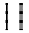
Low intensity

<---------------


<---------------


<---------------


The smoothing of these two different images, introducing PVE, results practically in the same image.

MQ 4-8-2003
<--- Previous
Next --->